Identifying Skin Rashes That May Indicate Skin Cancer
Skin rashes are common and often harmless, but some may signal underlying skin cancer concerns. Understanding the difference between typical skin irritations and potentially dangerous lesions can be crucial for early detection and treatment. While most rashes result from allergies, infections, or environmental factors, certain characteristics may warrant immediate medical attention. Learning to recognize warning signs helps distinguish between benign skin conditions and those requiring professional evaluation.

Early detection of skin cancer significantly improves treatment outcomes, making it essential to understand which skin changes require medical evaluation. Many people dismiss unusual skin appearances as simple rashes or age-related changes, potentially missing critical warning signs. Healthcare professionals emphasize that while most skin irregularities are benign, certain characteristics should prompt immediate consultation with a dermatologist.
Rashes That Look Harmless But Are Actually Skin Cancer
Several types of skin cancer can initially appear as innocent rashes or minor skin irritations. Basal cell carcinoma may present as a persistent red patch that resembles eczema or dermatitis. These patches often appear on sun-exposed areas like the face, neck, or hands and may occasionally bleed or form a crust. Unlike typical rashes that heal within days or weeks, cancerous lesions persist and may gradually change in appearance.
Squamous cell carcinoma can manifest as a rough, scaly patch that feels similar to dry skin or a minor abrasion. These areas may develop a raised, warty appearance or form open sores that fail to heal properly. The texture often feels different from surrounding skin, appearing thicker or more rigid than normal tissue.
Melanoma, the most serious form of skin cancer, can develop within existing moles or appear as new, unusual spots. Early-stage melanomas may look like irregular freckles or small, dark patches that seem relatively harmless. However, they typically exhibit asymmetry, irregular borders, color variations, or diameters larger than a pencil eraser.
Skin Cancer Pictures and Visual Recognition
Visual identification plays a crucial role in early skin cancer detection. Medical professionals recommend using the ABCDE method when examining suspicious spots: Asymmetry, Border irregularity, Color variation, Diameter changes, and Evolution over time. Photographs can help track changes in suspicious lesions, providing valuable documentation for healthcare providers.
Basal cell carcinomas often appear as pearly or waxy bumps, sometimes with visible blood vessels. They may also present as flat, flesh-colored or brown scar-like areas. Squamous cell carcinomas typically show as firm, red nodules or flat lesions with scaly, crusted surfaces. Advanced cases may develop into open sores with raised edges.
Melanomas display the greatest variety in appearance, ranging from black or brown spots to red, pink, white, or blue lesions. Some melanomas lack pigmentation entirely, appearing as pink or flesh-colored bumps. The key distinguishing factor is usually irregular features that differ from other moles or spots on the body.
Identifying Skin Rashes That May Indicate Skin Cancer
Proper identification requires systematic observation of skin changes over time. Normal rashes typically respond to treatment, appear symmetrical, and have consistent coloring throughout. Concerning lesions often fail to heal, continue growing, or develop new symptoms like itching, bleeding, or pain.
Location matters significantly in assessment. Skin cancers frequently develop on areas with high sun exposure, including the face, ears, neck, arms, and legs. However, they can occur anywhere on the body, including areas rarely exposed to sunlight like the palms, soles of feet, or under fingernails.
Texture changes provide important clues about potentially cancerous lesions. Healthy skin maintains consistent texture and flexibility, while cancerous areas may feel harder, rougher, or more rigid than surrounding tissue. Some lesions develop a waxy or translucent appearance that differs markedly from normal skin.
| Provider Type | Services Offered | Key Features |
|---|---|---|
| Dermatologists | Comprehensive skin cancer screening | Specialized training, dermoscopy equipment |
| Primary Care Physicians | Initial evaluation and referrals | Accessible first-line assessment |
| Dermatopathologists | Biopsy analysis and diagnosis | Laboratory expertise for tissue examination |
| Oncologists | Advanced cancer treatment | Specialized care for confirmed cases |
| Plastic Surgeons | Reconstructive procedures | Post-treatment cosmetic restoration |
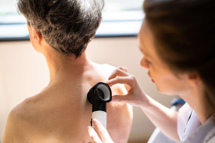
Regular self-examinations combined with professional screenings create the most effective detection strategy. Monthly self-checks should include examining all skin surfaces using mirrors for hard-to-see areas. Annual professional screenings are recommended for most adults, with more frequent evaluations for high-risk individuals.
Documentation proves invaluable for tracking changes over time. Photographing suspicious lesions provides objective records that help healthcare providers assess progression. Many smartphone applications now offer features specifically designed for skin monitoring, though they should supplement rather than replace professional medical evaluation.
Understanding personal risk factors helps determine appropriate screening frequency. Fair skin, family history of skin cancer, extensive sun exposure, history of severe sunburns, and numerous moles all increase skin cancer risk. Individuals with these factors should maintain heightened awareness and seek professional evaluation more frequently.
Recognizing potentially cancerous skin changes requires attention to detail and willingness to seek professional evaluation when concerns arise. While most skin irregularities prove benign, early detection of actual skin cancer dramatically improves treatment success rates. Regular monitoring, combined with prompt medical consultation for suspicious changes, provides the best protection against serious skin cancer complications.
This article is for informational purposes only and should not be considered medical advice. Please consult a qualified healthcare professional for personalized guidance and treatment.


