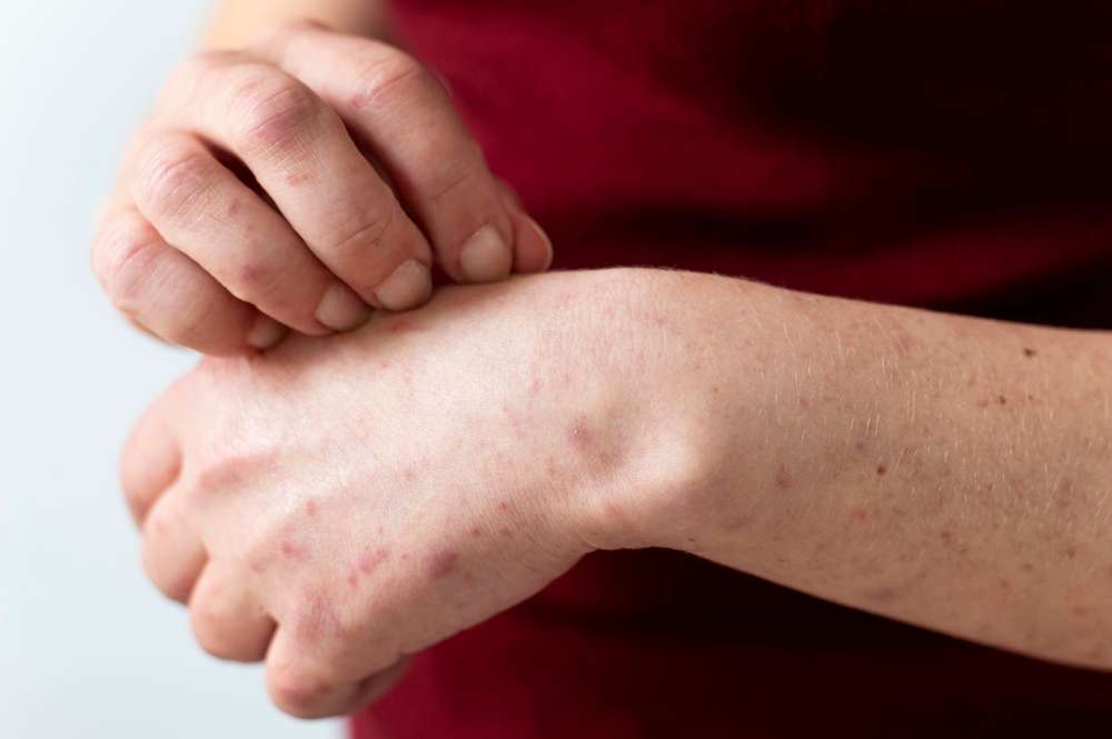
Mycosis Fungoides Visual Symptom Guide
Mycosis fungoides is a rare type of cutaneous T-cell lymphoma that primarily affects the skin. Understanding what this condition looks like at various stages can help individuals recognize symptoms early and seek appropriate medical evaluation. This guide explores the visual characteristics of mycosis fungoides, from initial presentation through more advanced stages, helping you identify key features that distinguish this condition from other skin disorders.

Mycosis fungoides presents unique visual characteristics that evolve over time, making early recognition challenging but crucial for timely intervention. This rare form of cutaneous lymphoma typically progresses through distinct stages, each with recognizable skin changes that can aid in identification and diagnosis.
What Does Mycosis Fungoides Look Like
Mycosis fungoides typically manifests as patches, plaques, or tumors on the skin, depending on the stage of progression. In its earliest form, the condition often appears as flat, scaly patches that may resemble eczema, psoriasis, or other common dermatological conditions. These patches are usually red or salmon-colored and can appear on areas of the body that receive less sun exposure, such as the buttocks, hips, and lower abdomen. The patches may be slightly itchy and can persist for months or even years before progressing. As the disease advances, the patches may thicken into raised plaques that have more defined borders and a deeper red or purple coloration. In later stages, tumors or nodules may develop, appearing as raised, dome-shaped lesions that can ulcerate or become infected. The visual presentation varies significantly between individuals, making clinical evaluation and biopsy essential for accurate diagnosis.
What Mycosis Fungoides Rash Looks Like in Early Stages
During the early stages, mycosis fungoides often mimics benign skin conditions, which contributes to delayed diagnosis. The initial rash typically appears as thin, flat patches that may be slightly scaly or dry to the touch. These patches often have irregular borders and can vary in size from a few centimeters to covering large areas of skin. The color ranges from light pink to deep red, and the affected areas may appear lighter or darker than surrounding skin. Early-stage lesions commonly occur on the trunk, particularly on areas covered by clothing, though they can appear anywhere on the body. The patches may come and go, leading some individuals to dismiss them as allergic reactions or temporary skin irritations. Itching is a common symptom in early mycosis fungoides, ranging from mild to severe, and may be the most bothersome aspect for many patients. The subtle nature of these early visual signs underscores the importance of dermatological evaluation when skin changes persist despite standard treatments for common conditions.
Recognizing Progressive Visual Changes
As mycosis fungoides progresses from the patch stage to the plaque stage, visual changes become more pronounced. Plaques appear as thickened, raised areas with well-defined edges and a more intense coloration than early patches. The surface may become rough or scaly, and the plaques can merge together to form larger affected areas. Some plaques develop a characteristic appearance with central clearing, creating a ring-like pattern. The skin may also develop changes in pigmentation, with some areas becoming hyperpigmented (darker) while others become hypopigmented (lighter). In advanced stages, tumors emerge as firm, raised nodules that can grow rapidly and may break down, forming open sores. These tumors can appear on previously affected areas or on previously normal skin. The visual progression helps clinicians stage the disease and determine appropriate treatment strategies.
Understanding Mycosis Fungoides Rash Pictures
Medical images and photographs of mycosis fungoides serve as valuable educational tools for both patients and healthcare providers. These visual references typically show the characteristic features at different stages, including the thin, scaly patches of early disease, the thicker, raised plaques of intermediate stages, and the nodular tumors of advanced disease. Dermatological atlases and medical literature contain extensive photographic documentation that illustrates the range of presentations across different skin types and stages. When reviewing such images, it is important to note that mycosis fungoides can appear differently on various skin tones, with color variations that may be more subtle on darker skin. Professional medical photography often includes close-up views showing texture and scale, as well as wider shots demonstrating distribution patterns. While these images are helpful for general awareness, they should never replace professional medical evaluation, as many skin conditions share similar visual features.
Distinguishing Features from Other Conditions
Several visual characteristics can help differentiate mycosis fungoides from other skin conditions, though definitive diagnosis requires biopsy. Unlike eczema, which typically responds well to topical corticosteroids, mycosis fungoides patches persist or worsen despite standard treatments. The distribution pattern often differs from psoriasis, which commonly affects extensor surfaces like elbows and knees, whereas mycosis fungoides favors sun-protected areas. The patches in mycosis fungoides may have a characteristic wrinkled or cigarette paper-like appearance that is less common in other conditions. Additionally, the progressive nature of the lesions, with transformation from patches to plaques to tumors over time, is distinctive. The presence of multiple stages simultaneously, such as patches and plaques coexisting, can also suggest mycosis fungoides. However, visual assessment alone is insufficient, and histopathological examination of skin biopsies remains the gold standard for diagnosis.
When to Seek Medical Evaluation
Recognizing when visual skin changes warrant professional evaluation is crucial for early detection and management. Individuals should consult a dermatologist if they notice persistent patches or plaques that do not respond to over-the-counter treatments within a few weeks. Skin changes that progressively worsen, spread to new areas, or develop new characteristics such as thickening or nodule formation require prompt assessment. Severe itching that interferes with daily activities or sleep, especially when accompanied by visible skin changes, should not be ignored. Any skin lesion that ulcerates, bleeds, or becomes infected needs immediate medical attention. Given the rarity of mycosis fungoides and its similarity to common conditions, multiple evaluations and biopsies may be necessary before reaching a definitive diagnosis. Early detection through awareness of visual symptoms can significantly impact treatment outcomes and quality of life.
Understanding the visual presentation of mycosis fungoides empowers individuals to recognize potential warning signs and seek timely medical evaluation. While this guide provides general information about appearance and progression, each case is unique, and professional dermatological assessment remains essential for accurate diagnosis and appropriate management of this complex condition.
This article is for informational purposes only and should not be considered medical advice. Please consult a qualified healthcare professional for personalized guidance and treatment.


